
Figure 6.
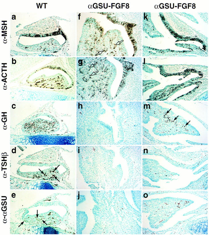
FGF8 promotes proliferation and opposes BMP activity in the pituitary gland. Phenotypic appearance of the pituitary gland in αGSU–FGF8 transgenic embryos at E17.5. Two transgenic embryos are shown, because the phenotype became more severe with higher copy number of the transgene [αGSU–FGF8 (f–j) strong phenotype, αGSU–FGF8 (k–o) weaker phenotype]. In both cases the gland exhibited striking dysmorphogenesis and increased cellularity (all photomicrographs taken at the same magnification). In αGSU–FGF8 (f,g), new clusters of lumenal-like cells have budded off (open arrows). Immunohistochemical analysis for the trophic hormones MSH (a,f,k), ACTH (b,g,l), GH (c,h,m), TSHβ (d,i,n), and αGSU (e,j,o) are shown, with selective loss of GH (h), TSHβ (i), and αGSU (j) in the case of the more severe phenotype and TSHβ (n) and αGSU (o) in the case of the weaker phenotype. The arrows mark clusters of positive cells for the respective hormones analyzed. Note that the dark dots, especially seen in the α–αGSU analysis of the αGSU–FGF8 (o) transgene, represent blood cells and not positive staining. Seven independent transgenic embryos at this stage were analyzed, with three showing the more severe abnormal phenotype then the other four.