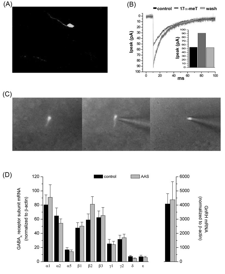
Figure 1.
Properties of GnRH Neurones. (A) Representative photomicrograph of a GnRH neurone from the GFP-GnRH transgenic line generously provided by SM Moenter (University of Michigan; [95]). (B) Acute modulation of GABAA receptor-mediated sPSCs in a GnRH neurone by 17α-methyltestosterone. Average currents (>30 neurones under each condition) recorded in aCSF alone (control), in aCSF supplemented with 1 µM 17α-methyltestosterone (17α-MeT) and following return to aCSF (wash). (C) Photomicrographs illustrating harvesting of the contents of a fluorescent GFP-GnRH neurone for single cell PCR analysis. (D) GABAA receptor subunit mRNA levels in GnRH neurones of gonadally-intact oil-injected (control) and AAS-treated male mice. Data are presented as the 2−ΔCT values, which indicate the average levels (relative to the housekeeping gene β-actin) of subunit mRNAs in GnRH neurones isolated from control (black; n= 10 mice) and AAS-treated (grey; n = 10 mice) for analysis of GABAA receptor subunit mRNA levels and from a separate cohort of 7 control and 7 AAS-treated mice for analysis of GnRH mRNA levels. Data in (D) are from Penatti et al. [69].

