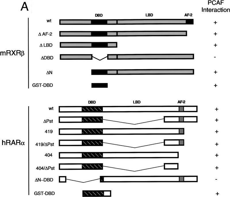
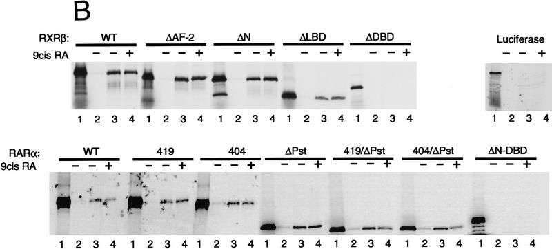
Figure 6.
Receptor domain analysis. (A) Diagram of full-length and truncated RXRβ and RARα and summary of PCAF binding (+, significant binding; −, no detectable binding). (B) 35S-Labeled receptors were incubated with rPCAF bound to anti-M2-Flag antibody conjugated to beads as in Fig. 4C. (Lane 1) Input (2 × 10⁴ cpm); (lane 2) control M2 beads without PCAF; (lanes 3,4) M2-rPCAF without (−) or with (+) 1 μm 9-cis RA, respectively. 35S-Labeled luciferase was tested as a negative control. (C) Control GST, GST–DBD from RARβ, or GST–DBD from RXRα (500 ng each) was incubated with rPCAF (+, 100 ng; ++, 300 ng) and PCAF binding was detected with anti-PCAF antibody. (Bottom) Loading of the control GST peptide, GST–DBD of RARα, and GST–DBD of RXRβ.


