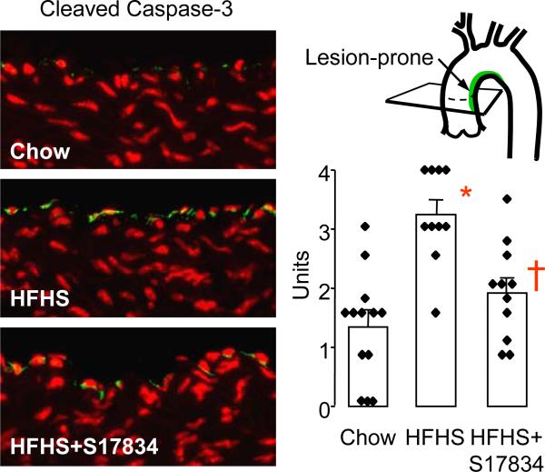
Figure 4.
S17834 prevents the increase in cleaved caspase-3 caused by HFHS diet in the lesion-prone aortic endothelium of C57BL6 wild type mice. Examples of immuno-fluorescent staining for cleaved caspase-3 in lesion-prone aortic endothelial cells are shown at left. The FITC-tagged secondary antibody was used to detect the cleaved caspase-3 antibody (green) in the endothelial cells. Nuclei were counterstained with propidium iodide (red). The bar graph at right shows the mean ± sem of the average scores obtained for each aortic cross section from each of 10-13 mice in each group. A statistically significant increase in staining for cleaved caspase-3 was caused by HFHS diet compared with chow (*P<0.05), and S17834 treatment resulted in a significant decrease compared with mice fed HFHS diet (†P<0.05).

