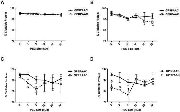
Figure 6. Percent Clottable Protein with varying PEG MW.
In all subfigures, the abscissa represents PEG size (kDa) with the 0 kDa mark indicating the associated peptide in non-PEGylated form. Percent clottable protein was quantified from the remaining protein in solution after clot formation. Data for GPSPAAC (●) and GPRPAAC (□) formulations at four molar ratios of fibrinogen:conjugate are displayed, (A) 10:1, (B) 1:1, (C) 1:10, and (D) 1:100. (Mean ± SD; n= 6–9; # denotes p < 0.05, * denotes p < 0.01, and $ denotes p < 0.001).

