
Figure 6.
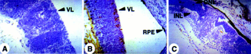
IRBP-expressing Rb−/−;p107−/− cells disappear from the retina before P15. Immunohistochemical staining for p53DD protein with pAb421 plus counterstaining with hematoxilyn. (A) No staining in the dysplastic retina of a chimeric Rb−/−;p107−/− embryo (E17.5). (B) Retina of a chimeric Rb−/−;p107−/−;hIRBPp53DD embryo (E17.5) with positive cells in the ventricular layer (arrows). (C) No staining in the retina with malignant nodular growth of a young chimeric Rb−/−;p107−/−;hIRBPp53DD mouse (P15). (RPE) Retinal pigment epithelium; (VL) ventricular layer; (ONL) outer nuclear layer; (INL) inner nuclear layer. Magnification (A,B) 400×; (C) 200×.