
Fig. 3.
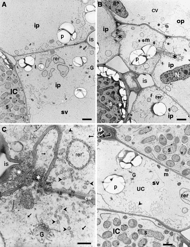
Shrinkage of vacuoles and cell wall infoldings in the inner parenchyma of a 2-week-old pea root nodules observed after shoot removal. (a) Shrinking vacuoles and diluted cytoplasm in the inner parenchyma cells. RER rings are located close to the radial cell walls. (b) Cell wall infoldings are visible on radial cell walls. (c) Structure of the cell wall infoldings. Notice fine, fibrillar material (arrows) and numerous vesicles (arrowheads) visible in the vicinity of the infoldings. Stars indicate rer profiles which fill niches formed by folded cell walls. (d) Vacuole shrinkage is visible in uninfected cells after shoot removal. Abbreviations: arrowhead—small vesicles, cw—cell wall, CV—shrinking vacuole, G—Golgi body, IC—infected cell, is—intercellular space, ip—inner parenchyma, op—outer parenchyma, m—mitochondrium, p—plastid, rer—rough endoplasmic reticulum, s—symbiosome, small arrows—fibrillar material, star—electron-opaque material within cell wall infolding, UC—uninfected cell. Bars: A and B = 2 μm, C = 0,5 μm, D = 1 μm