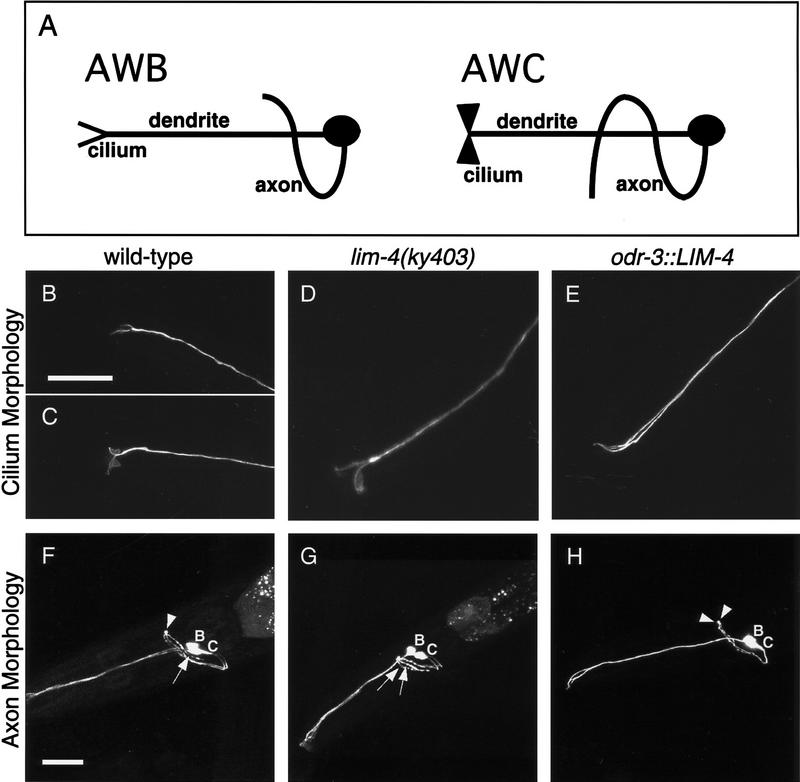
Figure 4.
LIM-4 controls the morphology of sensory cilia and axons. (A) Diagrams illustrating the wild-type morphologies of AWB and AWC neurons. Cilia are specialized sensory structures at the tips of dendrites. An AWB cilium is a simple two-pronged structure, resembling a tuning fork, whereas an AWC cilium is more membranous or fan-like. The U-shaped AWB axon extends into the nerve ring and stops at the dorsal midline. The S-shaped AWC axon continues past the dorsal midline to a ventral position on the contralateral side of the nerve ring. (B–E) Confocal Z-series projections of AWC and AWB cilia. Wild-type cilium morphologies of AWB (B) and AWC (C), visualized with str-1::GFP and str-2::GFP, respectively. (D) An AWB cilium in a lim-4(ky403) mutant, visualized with str-2::GFP, resembles the fan-like AWC cilia. (E) An odr-3::LIM-4-expressing animal visualized with str-1::GFP; both cilia exhibit the simple tuning fork morphology of AWB. (F–H) Confocal Z-series projections of GFP-expressing AWC and AWB neurons. Mosaic animals in which the neurons on only one side of the animal expressed GFP were chosen to simplify analysis of axon trajectories. (F) In animals with wild-type axon morphologies, the AWB axon stops at the ventral midline (arrowhead), whereas the AWC axon continues ventrally past the midline (arrow). This picture is of a lim-4 mutant expressing str-2::GFP displaying the wild-type axon morphologies for AWB and AWC. Wild-type animals expressing str-1::GFP and str-2::GFP exhibited an identical phenotype 100% of the time (Table 2). (G) A lim-4(ky403) mutant expressing str-2::GFP; both AWB and AWC axons exhibit the longer S-shaped AWC axon morphology (arrows). (H) An odr-3::LIM-4-expressing animals with str-1::GFP; both AWB and AWC axons stop at the midline (arrowheads). Quantitative analysis of these phenotypes is presented in Table 2. Anterior is left and dorsal is up in all panels. Scale bars, 20 μm. Bar in B applies to B–E; bar in F applies to F–H.

