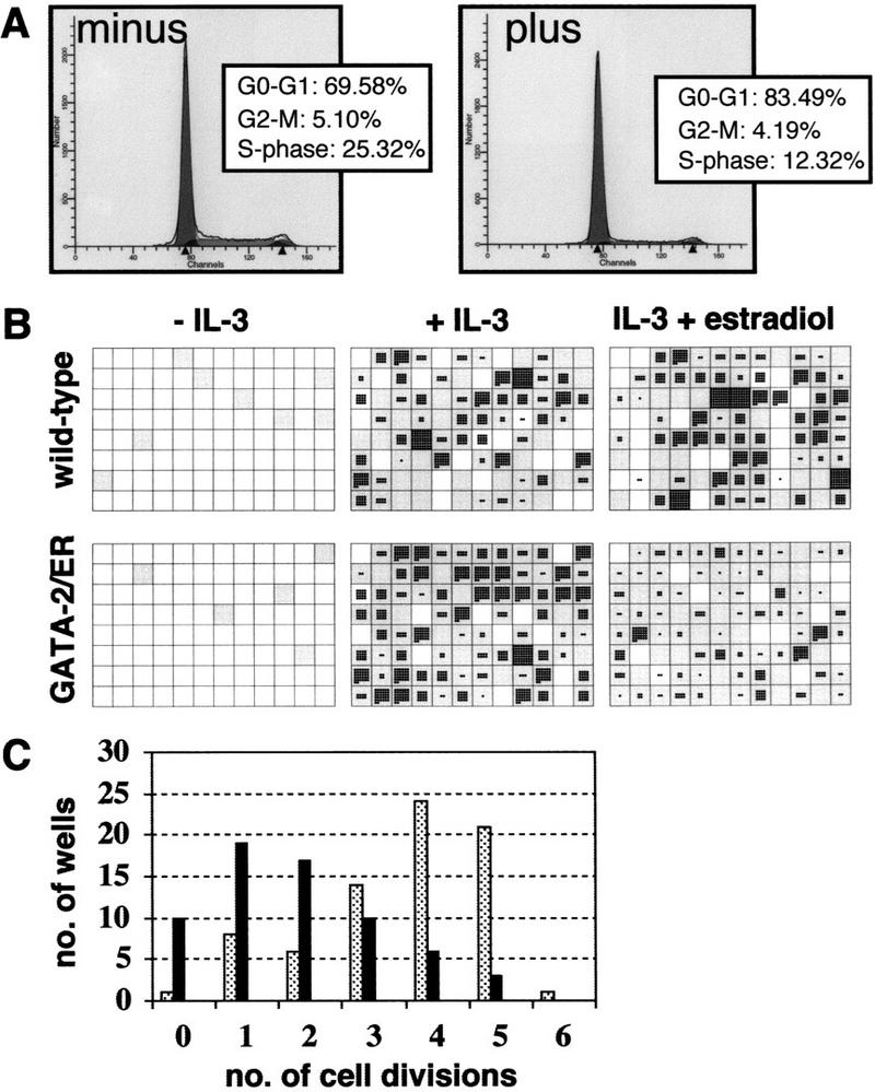
Figure 2.

Analysis of GATA-2-mediated growth arrest. (A) cell cycle analysis of GATA-2/ER cells. GATA-2/ER cells exponentially growing in IL-3-containing medium were incubated minus (left) or plus (right) 0.2 μm β-estradiol for 48 hr. The cells were harvested and stained with Hoechst dye, and cell cycle distribution analyzed on a Becton-Dickinson FACS Vantage flow cytometer using MODFIT LT v. 2.0 software (Verity Software, USA). The plot shows a typical result obtained with A7 GATA-2/ER no. 1. Similar results were obtained with other clones, and each experiment was performed at least three times. (B) A7 GATA-2/ER no. 1 cells were seeded into 96-well plates in the absence of IL-3 or in IL-3 (10 ng/ml) with or without 0.2 μm β-estradiol. The number of cells was counted at 24 hr and 3 days. For each experiment two trays per condition were assessed, and the experiment repeated three times. A typical set of plates is shown. Wells that contained a viable cell at 24 hr are shown in gray. The number of cells in each well after 3 days growth is shown, with each cell represented by a single black dot. (C) Summary of the number of GATA-2/ER cell divisions that had occurred in each well in the presence of IL-3 but absence (stippled bars) or presence (solid bars) of β-estradiol. The results represent a count of the number of wells from one experiment but are typical of three independent experiments.
