
Figure 3.
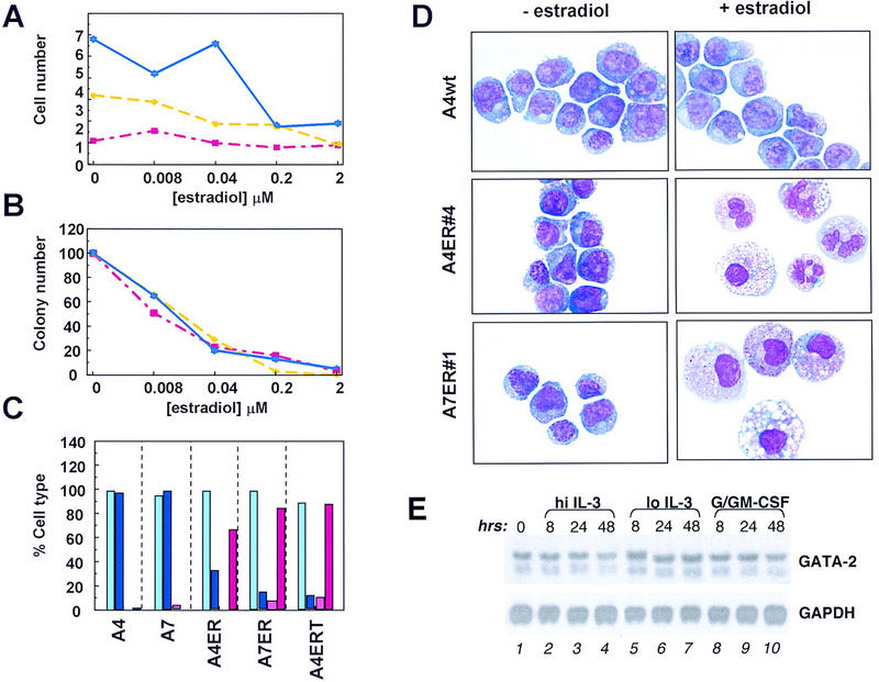
Enforced GATA-2 expression leads to differentiation. (A) A7 GATA-2/ER no. 1 cells were seeded at 8 × 10⁴ cells/ml in IL-3-containing medium in the presence of various concentrations of β-estradiol. Cells were harvested after incubation at 37°C for 1, 2, or 3 days. The number of viable cells was determined by trypan blue staining and plotted against the concentration of β-estradiol used. The experiment was performed three times, and the plot shows the results from a typical experiment. Day 1, 2, and 3 samples are plotted in red, yellow and blue, respectively. (B) A7 GATA-2/ER no. 1 cells were treated for 1, 2, or 3 days with β-estradiol as described above. After harvesting the cells, washing twice with PBS, 2 × 10³ cells/ml were plated in IL-3-stimulated soft agar CFC assays. Plates were incubated at 37°C for 7 days, and the number of colonies counted and expressed as a percentage of the untreated control. The experiments were performed in parallel to those in A, and the plot shows the results from a typical experiment. Day 1, 2, and 3 samples are plotted in red, yellow, and blue, respectively. (C) Wild-type FDCP-mix A4 and A7 cells and clones of A4 and A7 expressing GATA-2/ER or GATA-2/ERT were grown in IL-3-containing medium for 3 days in the absence or presence of the appropriate inducer (0.2 μm β-estradiol or 2 μm tamoxifen). The cells were harvested, cytospun, and stained. The morphology was determined as either blast cells or granulocyte/macrophage cells, and the results expressed graphically as a percentage of the total. Light blue, blasts (− estradiol); dark blue, blasts (+ estradiol); pink, granulocytes/macrophages (− estradiol); red, granulocytes/macrophages (+ estradiol). (D) Wild-type and GATA-2/ER-expressing FDCP-mix clones were treated as described for C. In the absence of β-estradiol (− estradiol) all of the cells had a primitive morphology characteristic of blast cells. Incubation of control cells (A4wt) in the presence of β-estradiol (+ estradiol) did not change their morphology. However, clones containing inducible GATA-2 differentiated and showed up-regulation of the myeloid surface markers Mac-1 and GR-2 (not shown). The morphologies observed were characteristic of either granulocyte/monocyte (A4 GATA-2/ER no. 4) or monocytes (A7 GATA-2/ER no. 1). Granulocytes have a condensed segmented nucleus with a pale staining cytoplasm containing some granules, whereas monocytes are larger cells with a large cytoplasmic/nuclear ratio and a slightly kidney-shaped nucleus. The morphology of these cells is typical of mature murine hematopoietic cells differentiated in vitro. Typically, some vacuolation of both immature and mature cells may be seen in these in vitro culture conditions. (E) The level of GATA-2 mRNA in wild-type FDCP-mix cells during granulomonocytic differentiation was assessed by Northern blot using a portion of the murine GATA-2 cDNA as a probe. FDCP-mix cells were cultured for the time periods indicated (0, 8, 24, 48 hr) under normal self-renewal conditions (hi IL-3, lanes 2–4), the low IL-3 condition normally used during the G/GM-CSF culture (lo IL-3, lanes 5–7), or granulomonocytic differentiation conditions (G/GM-CSF, lanes 8–10). The GATA-2 mRNA is detected as two bands of 3.5 and 2.9 kb. Equal RNA loading in each lane was confirmed by stripping the membrane and reprobing for GAPDH mRNA (1.2 kb).