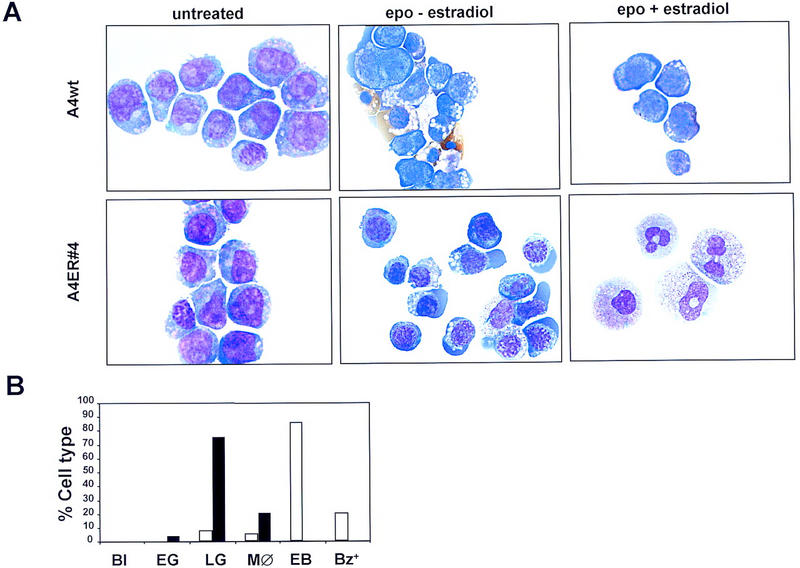
Figure 5.

Erythroid potential of wild-type and GATA-2/ER FDCP-mix cells. (A) FDCP-mix clones were differentiated in the presence of EPO (5 U/ml), hemin (0.2 mm), and low IL-3 (0.05 ng/ml). The morphology of differentiated cells is described in the legend to Fig. 3D; erythroid cells are characterized by their size being smaller than blast cells, a basophilic cytoplasm, and nuclei containing condensed chromatin. (Top) Typical photomicrographs of wild-type FDCP-mix A4 cells, either untreated (no EPO or β-estradiol), differentiated in the presence of erythroid differentiation factors (epo) but the absence of β-estradiol, or differentiated in the presence of epo and β-estradiol. (Bottom) The morphology of the A4 GATA-2/ER no. 4 cells cultured under the same conditions. (B) Summary of the number of each cell type expressed as a percentage of the total cell number for A4 GATA-2/ER no. 4 cells in the presence of EPO and absence (open bars) or presence (solid bars) of β-estradiol. (Bl) Blasts; (EG) early granulocytes; (LG) late granulocytes; (MØ) monocytes; (EB) erythroblasts; (Bz+) benzidine-positive cells.
