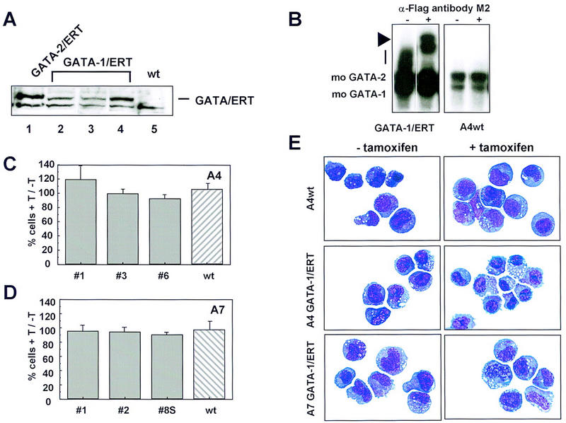
Figure 6.

Enforced expression of GATA-1/ERT in FDCP-mix A4 and A7 cells. (A) FDCP-mix clones expressing GATA-1/ERT (lanes 2-4) were identified by Western blotting using the M2 anti-Flag antibody. Comparison with a high expressing BA/F3 GATA-2/ERT clone (lane 1) shows that the expression levels obtained for GATA-1/ERT and GATA-2/ERT are similar. Untransfected FDCP-mix A4 (wt) was included (lane 5) as a control for the specificity of the antibody. (B) The expression level and DNA-binding activity of GATA-1/ ERT was assessed by EMSA as for GATA-2/ERT. The GATA-1/ERT protein DNA complex migrates as a smear (vertical bar), which is clearly absent from wild-type FDCP-mix A4 nuclear extract. Its identity was confirmed by using the M2 anti-Flag antibody (+ lanes) to supershift the complex (arrowhead). (C) The growth of wild-type (hatched bar) and three GATA-1/ERT-expressing (shaded bars) FDCP-mix A4 clones (nos. 1, 2, and 3) in the presence of 2 μm tamoxifen (T) is shown expressed as a percentage of their growth in the absence of tamoxifen. The results are the average of three experiments ±s.e.m.. (D) The experiment described in C was repeated for wild-type FDCP-mix A7 (hatched bar) and three GATA-1/ERT-expressing (shaded bars) FDCP-mix A7 clones. The results are essentially identical to those obtained in FDCP-mix A4 and are the average of three experiments ±s.e.m.. (E) Morphological analysis of cells expressing GATA-1/ERT. Cells were cultured in the absence (− tamoxifen) or presence (+ tamoxifen) of 2 μm tamoxifen for 3 days, harvested, cytospun, and stained. The cells used were A4 GATA-1/ERT clone 3, A7 GATA-1/ERT clone 2, and wild-type FDCP-mix A4. All cells have an essentially blast-like morphology both in the absence and presence of tamoxifen.
