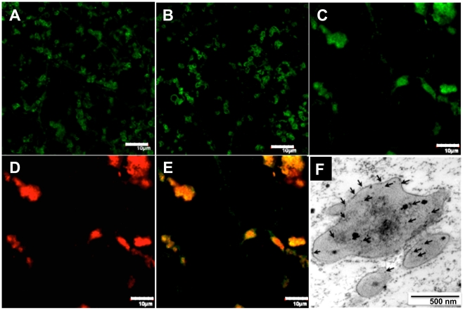
Figure 4. Co-localization of EiP and prolamins in the irregular protein bodies.
(A–C) Prolamins labeling with FITC fluorescence in WT (A), SP (B) and SA (C) endosperm cells. (D) ELP labeling with Rhodamine Red fluorescence in SA endosperm cells. (E) Merged pictures of (C) and (D) Bars, 10 µm. (F) Immuno-gold labeling with anti-prolamin antibody confirmed the deposition of prolamins in the iPBs (indicated by arrows). Bar, 500 nm.

