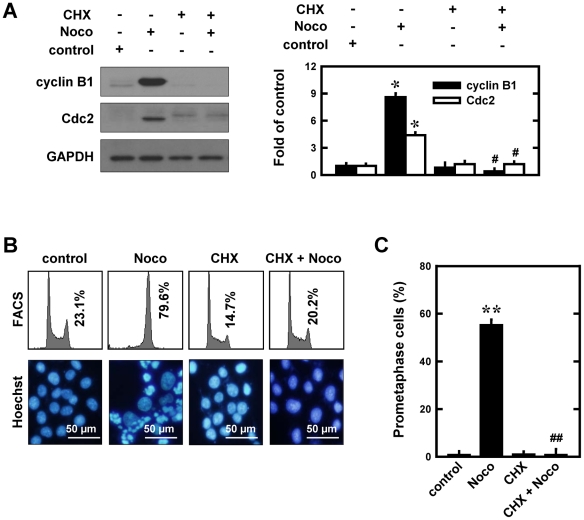
Figure 6. Effect of cycloheximide (CHX) on nocodazole-induced prometaphase arrest in MCF-7 cells.
A (left panel). Cells were pre-treated for 2 h with cycloheximide (5 µg/mL) and then stimulated for additional 12 h with 250 nM nocodazole. Total cell lysates were analyzed by Western immunoblotting for cyclin B1 and Cdc2. A (right panel). The relative protein levels of cyclin B1 and Cdc2 are calculated according to their densitometry readings, which are normalized according to the corresponding readings for the GAPDH protein bands. Each value is mean ± S.D. from triplicate measurements. * P<0.05 versus vehicle-treated control; # P<0.05 versus nocodazole treatment. B (upper panel). Cells were pre-treated for 2 h with cycloheximiden (5 µg/mL) and then stimulated for additional 12 h with 250 nM nocodazole. The DNA content of cells was analyzed using flow cytometry as described in the Material and Methods section. B (lower panel). Nuclei were stained with Hoechst-33342, and examined using a fluorescence microscope for prometaphase cells (original magnification, ×200). C. Quantitative data on prometaphase-arrested cells. Each bar is a mean ± S.D. value from three separate experiments. ** P<0.01 versus the vehicle-treated control; ## P<0.01 versus nocodazole treatment.

