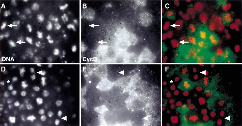
Figure 7.
Low levels of PIMdba–myc expression allow sister chromatid separation in pim mutants. pim1/ pim1 (A–C) or pim1/ pim1, gpimdba–myc (D–F) embryos at the stage of mitosis 15 were labeled with a DNA stain (DNA; A,D) and antibodies against Cyclin B (CycB; B,E). Regions of the dorsal epidermis are shown. In the merged panels (C,F) DNA labeling is shown in red and Cyclin B in green. Arrows in A–C indicate cells that have failed to separate sister chromatids even though they have progressed beyond the metaphase–anaphase transition as evidenced by lack of anti-Cyclin B labeling. Arrowheads in D–F indicate cells that have separated sister chromatids successfully after the metaphase–anaphase transition. (*, D–F) Cell with an anaphase bridge.

