
Figure 1.
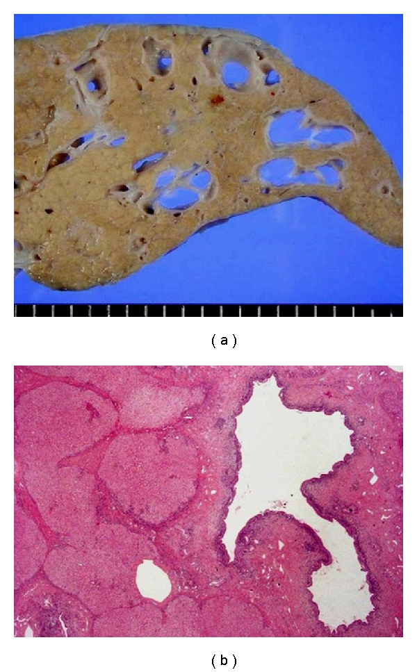
The liver of a Caroli's disease patient with CHF. Multiple cystic dilatations of the intrahepatic bile ducts are grossly (a) and histologically (b) visible. Hematoxylin-eosin staining (b).
Official websites use .gov
A
.gov website belongs to an official
government organization in the United States.
Secure .gov websites use HTTPS
A lock (
) or https:// means you've safely
connected to the .gov website. Share sensitive
information only on official, secure websites.
The liver of a Caroli's disease patient with CHF. Multiple cystic dilatations of the intrahepatic bile ducts are grossly (a) and histologically (b) visible. Hematoxylin-eosin staining (b).