Figure 4.

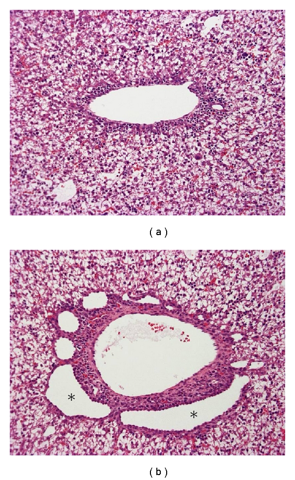
Ductal plate malformation in the fetal liver of a PCK rat. Compared with a normal fetal rat (a), dilatation of the ductal plate is evident in the PCK liver (b, asterisks). Hematoxylin-eosin staining (a, b).

Ductal plate malformation in the fetal liver of a PCK rat. Compared with a normal fetal rat (a), dilatation of the ductal plate is evident in the PCK liver (b, asterisks). Hematoxylin-eosin staining (a, b).