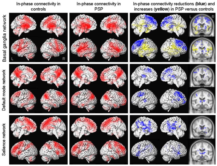
Figure 2.
Resting-state fMRI results from ICA-based analyses of the basal ganglia network, DMN and salience network. Left: Patterns of in-phase voxel-wise connectivity observed in control subjects; Middle: Patterns of in-phase voxel-wise connectivity observed in PSP subjects; Right: Patterns of reduced (shown in blue) and increased (shown in yellow) in-phase connectivity in the PSP subjects compared to controls, shown on both 3D renders and slices through the basal ganglia and thalamus. Results are shown after cluster-level correction for multiple comparisons at p<0.05.

