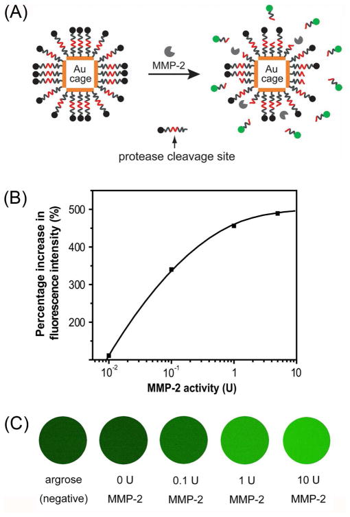
FIGURE 9.
(A) A schematic of the dual probe that could be activated by an enzyme. The probe is composed of a AuNC and fluorescent dyes linked together through a peptides cleavable by an enzyme. (B) Percentage increase in fluorescence intensity as a function of MMP-2 activity (20 fmol FITC-GKGPLGVRGC-AuNCs, incubation time: 12 h); (C) Fluorescence images of FITC-GKGPLGVRGC-AuNCs after incubation with different concentrations of MMP-2 at 37 °C for 12 h. The sample was mixed with 1.5% agarose to form gel for imaging.35

