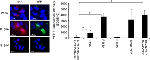
Figure 2.
Split YFP as a tool to image Bax conformational change. C-YFP–HA and N-YFP–HA were independently cloned downstream or upstream to wild-type Bax or different Bax mutants. MCF-7 cells were transiently transfected and 24 h later the levels of YFP were measured either using a fluorescence plate reader (right panel) or by fluorescence microscopy, and correlated with anti-HA staining (red, left panel, n=4; aP<0.05; bP<0.00001; cP<0.0005)

