Figure 5.
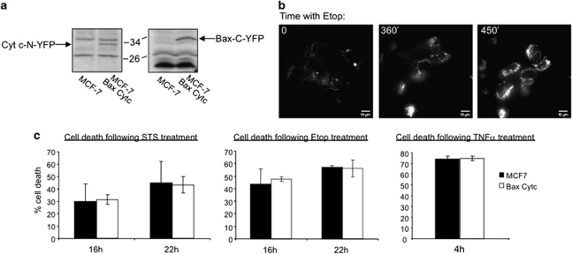
Stable expression of C-YFP–Bax and Cyt c–N-YFP. (a) MCF-7 cells were infected with lentiviruses containing C-YFP–HA–Bax and Cyt c–N-YFP. Stable clones were selected using blasticidine and the expression level was analyzed by Western blot using anti-Cyt c and anti-HA antibodies. (b) MCF-7 cells stably expressing C-YFP–HA–Bax and Cyt c–N-YFP were treated with etoposide (100 μM) and followed using the DeltaVision system. (c) A total of 2 × 10⁴ naive MCF-7 cells and cells that stably express C-YFP–HA–Bax and Cyt c–N-YFP were plated in black, 96-well plates and treated with staurosporine (1 μM, 16 and 22 h), etoposide (100 μM, 16 and 22 h) and TNFα with ActD (4 h). Cell viability was measured using the CellTiter Blue reagent and cell death was calculated relative to non-treated cells. No statistical significant differences were observed (P>0.1)

