Figure 2.
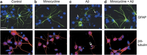
β-Amyloid (Aβ) induces morphological changes in astrocytes. Representative images from mixed cortical cultures treated for 48 h with (a) vehicle, (b) 20 μM minocycline, (c) 10 μM Aβ or (d) 10 μM Aβ with a 24 h pretreatment with 20 μM minocycline. Cells were fixed and immunostained with antibodies against glial fibrillary acidic protein (GFAP) (green), to label astrocytes, or βIII-tubulin (red), to label neurons. Bisbenzimide was used to stain nuclei (blue). Single arrows indicate astrocytic terminal swellings, and double arrows indicate apoptotic nuclei. Scale bar: 50 μm. N=9

