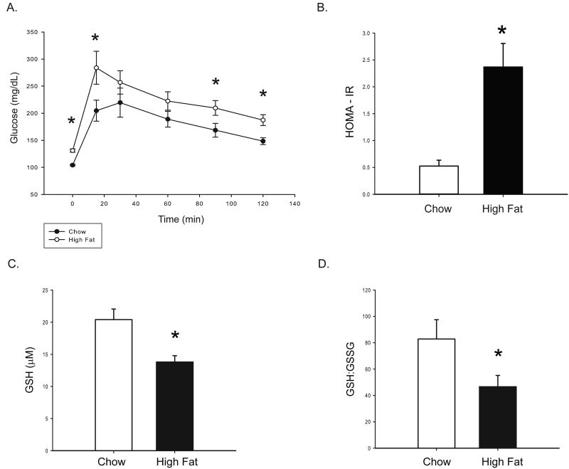
Figure 1. Peripheral glucose tolerance and oxidative stress.
After an overnight (12 hour) fast, an intraperitoneal glucose tolerance test was performed. A 60% glucose solution was administered intraperitoneally at 2g glucose/kg body weight. (A) Glucose was measured in tail blood at six time points: 0,15,45,60, 90, and 120 minutes after the glucose bolus (injection at t=0). Over the course of the test, glucose levels were higher in HF fed animals. HOMA-IR was also significantly increased (B), indicating insulin resistance. Serum levels of reduced glutathione (C) and the ratio of reduced to oxidized glutathione (D) were measured to gauge diet-induced oxidative stress. HF-feeding decreased reduced glutathione levels and decreased the ratio of reduced to oxidized glutathione. Values are means ± SE for 11-15 rats per group. *P<0.05 chow vs. HF.

