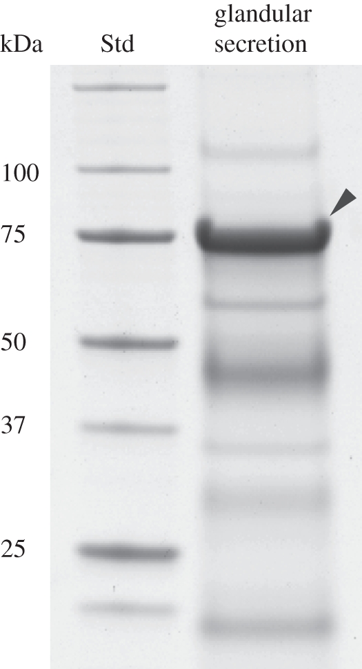
Figure 2.

Coomassie Brilliant Blue-stained one-dimensional SDS protein gel of the larval glandular secretion of P. vitellinae. The most prominent band marked with an arrow corresponds to SAO. The size of the protein standard on the left is indicated in 103 Da.
