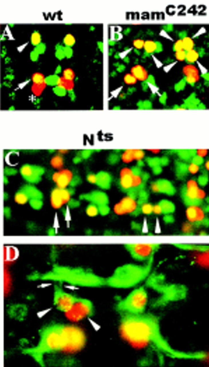
Figure 1.

Mutations in mam and N are associated with RP2sib to RP2 and pCC to aCC fate transformations. (A,B,C) Ventral views of stage 15 embryos double stained with rabbit anti-Eve (red) and mouse anti-Zfh-1 (green). (A,B) Anterior is up; (C) anterior is left. (A) wt RP2 (arrowhead) and aCC (large arrow), but not pCC (asterisk), coexpress Eve and Zfh-1 (yellow). (B) mamC242. (C) Nts1. Note the different sizes of the RP2 nuclei in B and C (arrowheads). The right hemisegment in B depicts four RP2 neurons that presumably derive from two parental NB4-2. (D) Ventral view of a stage 15 whole-mount Nts1 embryo stained with rabbit anti-Eve (red) and mouse-mAb 22C10 (green). Anterior is up. Note the ipsilateral axon projections of both RP2s (small arrows).
