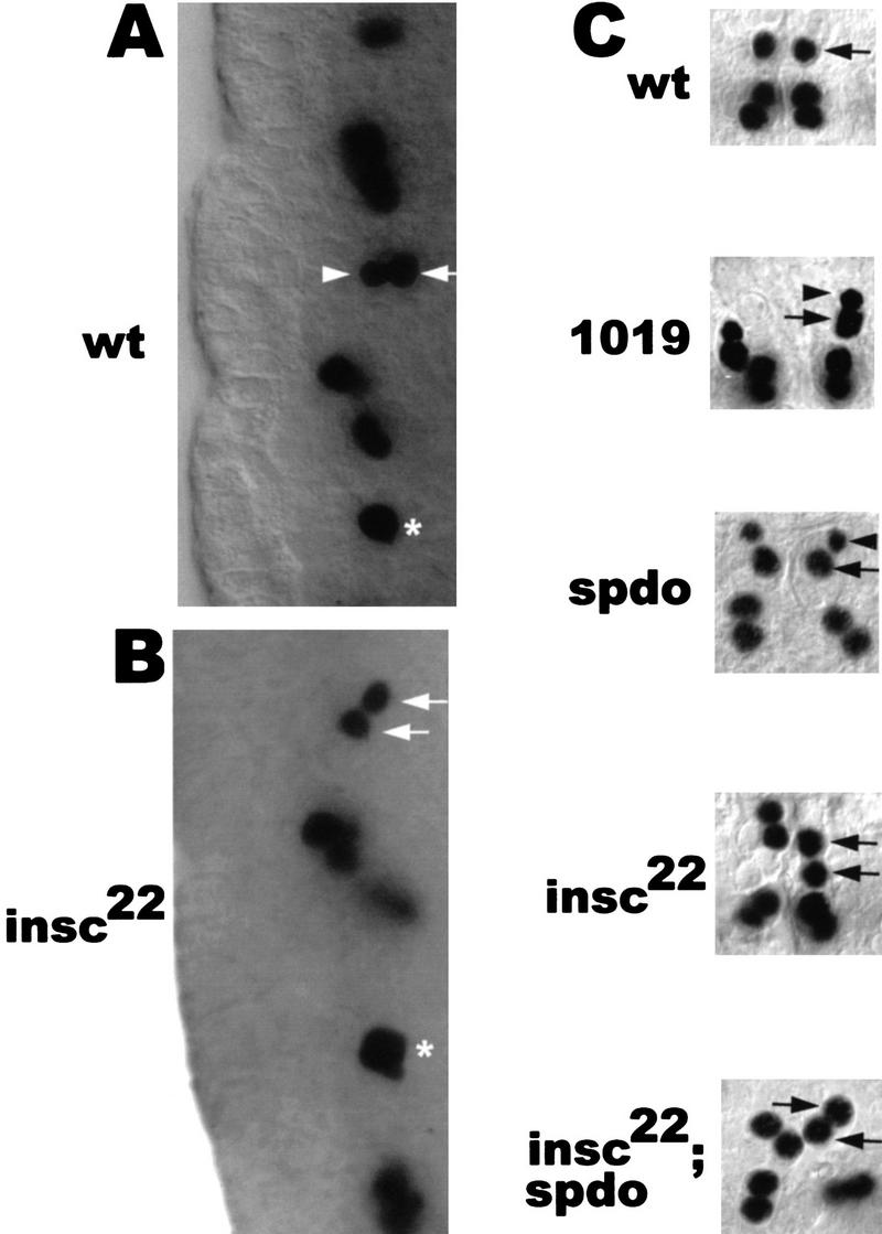
Figure 2.

Mutations in spdo result in RP2sib to RP2 fate transformation with respect to marker gene expression but not with respect to cell/nuclear size. (A,B) Lateral views of stage 11 embryos stained with anti-Eve. Ventral (apical) is to the left. (A) In wt, GMC4-2a divides into a smaller and a larger cell. The newborn siblings are oriented perpendicular to the apical surface with the larger cell (future RP2) in the more dorsal position. (B) In insc22, GMC4-2a divides into sibling cells of equal size that are rarely oriented perpendicular to the apical surface. (Arrows) RP2 neuron; (arrowheads) RP2sib; (asterisks) undivided GMC4-2a. (C) Dorsal views of dissected wild-type and various mutant stage 15 embryos stained with anti-Eve. Anterior is up. In GA1019 (1019) embryos, GMC4-2a does not undergo cytokinesis and binucleated cells with two Eve-expressing nuclei are formed. Note the difference in the sizes of the two nuclei (arrowhead vs. arrow) derived from GMC4-2a in spdo and GA1019 embryos.
