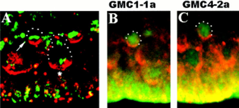
Figure 4.

Insc protein forms apical crescents in dividing GMCs. Lateral views of wt stage 11 embryos; basal (dorsal) is up. (A) Double staining with rabbit anti-Insc (red) and DNA stain (green). (Arrow) GMC in metaphase. Note that the metaphase plate is oriented horizontally with respect to the apical surface. (Asterisk) Dividing neuroblast. (B,C) GMC1-1a and GMC4-2a double stained with anti-Insc (red) and mouse anti-Eve (green).
