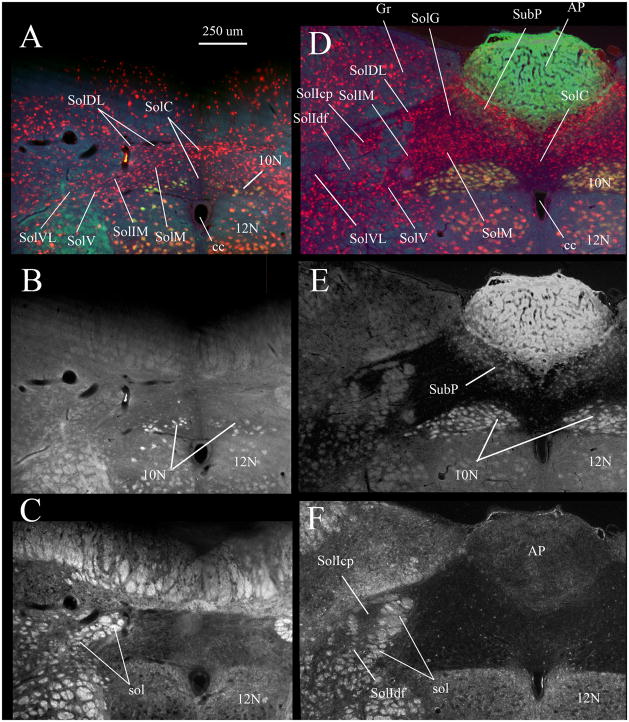
Fig. 1. Caudal NTS subdivisions viewed via NeuN immunostaining (A, D), FluoroGold labeling of vagal motoneurons and area postrema (B, E) and by darkfield illumination of solitary tract axons (C, F).
Nuclei composing the caudal NTS are indicated in A and D. A–C show the same section ~ 300 μm caudal to obex where the commissural nucleus of the solitary tract (SolC) is most prominent. D–F show a single section ~ 400 μm rostral to obex where the area postrema is near its maximal mediolateral extent.
In A and D red neurons are immunofluorescent for neuron specific nuclear protein (NeuN). In A, B, D and E labeling in the dorsal motor nucleus of the vagus (10N), hypoglossal nucleus (12N), area postrema (AP) and subpostrema nucleus (SubP) resulted from a subcutaneous injection of FluoroGold. In C and F darkfield illumination identifies the myelinated fascicles of the solitary tract (sol).
A and D are true color RGB images captured using a triple band-pass epifluorescence filter (uv/DAPI, FITC, Texas Red) and a RGB liquid crystal filter. Images in B and E were extracted from the green channel of the RGB images in A and D respectively.

