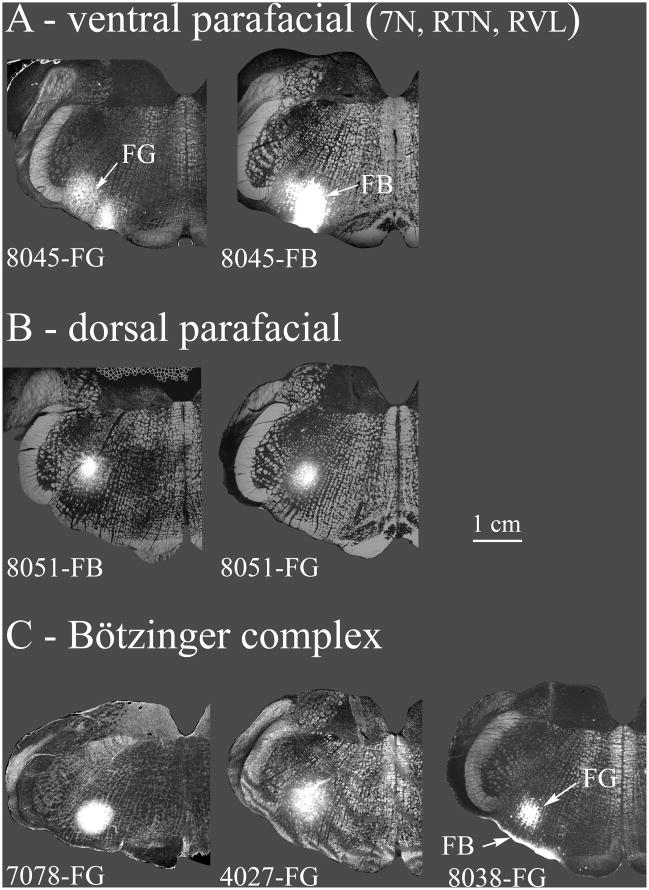
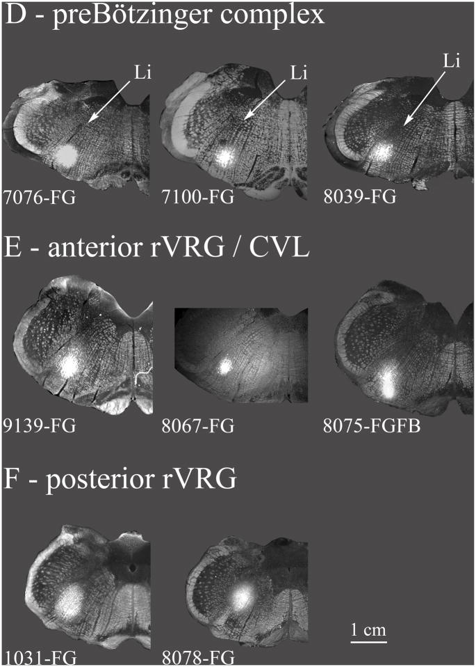
Fig. 3. Retrograde tracer injection sites.
The various retrograde tracer injections in the VRC and nearby areas (7N, RVL, CVL) are summarized. A: Ventral parafacial injections were located ventrally at the caudal end of the facial nucleus. These included the facial nucleus but also the RTN, and a rostral portion of RVL. These injections are depicted in greater detail in Fig. 4. B: Injections are located near the caudal facial nucleus but more dorsal than in A. The dorsal parafacial injections are depicted in greater detail in Fig. 5. C: Injections were classified as within the Bötzinger complex based on their location just caudal to the facial nucleus and by the types of neurons recorded proximal to the injections (see results). In case 8038 the bright band of labeling ventral to the FG injection resulted from a second injection tract where an FB injection was inadvertently made at the ventral surface of the medulla. Very few FB retrogradely labeled neurons were noted in the caudal NTS. Case 7078 is depicted in greater detail in Fig. 6. D: Injections were classified as within the preBötzinger complex on the basis of extracellular recordings near the tracer injection sites (see results) along with the location of the injection at the caudal end of the compact part of n. ambiguus, at approximately the same level as the folded portion of the linear nucleus of the medulla (Li) and just rostral to the lateral reticular formation. Case 7076 is depicted in greater detail in Fig. 7. E: Injections in the anterior part of rVRG (arVRG) were identified by the presence of inspiratory neurons at the injection site, the presence of the anterior portion of the lateral reticular nucleus and the absence of the area postrema; it was distinguished from posterior rVRG by the differential responses to DLH injection, bradypnea in anterior but not posterior rVRG. CVL neurons are generally located ventral to anterior rVRG (e.g. Figs. 2, 10) although functional overlap is indicated where DLH injections aimed at arVRG decrease blood pressure and heart rate (see results and case 9139 depicted in Fig. 8). F: Posterior rVRG injections, as with anterior rVRG, were localized by the presence of inspiratory neurons and the lateral reticular nucleus. In case 8075 FG and FB were differentially targeted at posterior VRG and CVL but overlapped due to the somewhat elongated shape of the FB injection. The presence of the area postrema helps discriminate the posterior rVRG from anterior rVRG as does the absence of significant changes in respiratory rhythm consequent to local DLH injections. Case 1031 is described in Fig. 9.
Injection sites are depicted using a grayscale darkfield image of the relevant section as the base image overlaid by the injection site image using the “lighten” command in Photoshop®. Injections were saved as epifluorescent images for native FG/FB fluorescence, or as inverted brightfield images for DAB stained FG sections.


