
Figure 1.
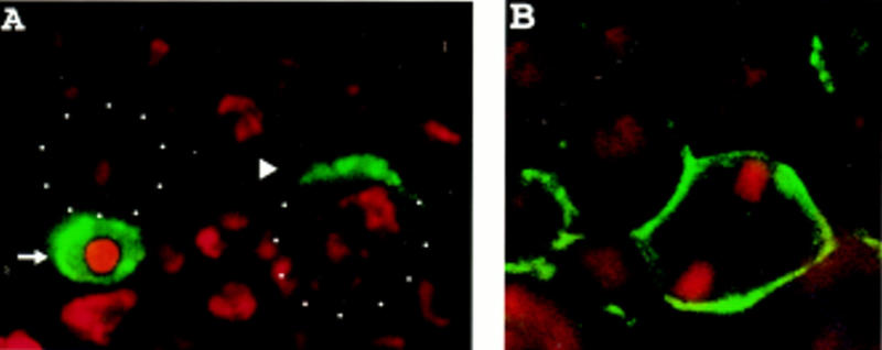
Microfilaments are required for the localization of Miranda to the apical then basal cortex. (A) Wild-type embryos were stained with anti-Miranda antibody (green) and propidium iodide to visualize DNA (red). Apical is toward the top; white dots indicate neuroblast cell borders in this and all subsequent figures. In the neuroblast at right, which is in early prophase, Miranda is localized to the apical cell cortex (arrowhead). The neuroblast at left has nearly completed mitosis. Miranda is present exclusively in the basal daughter cell (arrow). (B) Wild-type embryos were treated with 200 μm latrunculin A for 20 min and stained for Miranda (green) and DNA (red). Under these conditions, phalloidin reactivity is virtually abolished (Knoblich et al. 1997), and Miranda is localized along the entire cortex of mitotic neuroblasts.