Fig. 2.
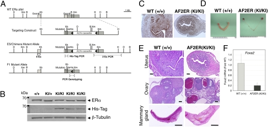
Targeting strategy and confirmation of L543A, L544A ERα knock-in mutation. (A) Schematic illustration of the targeting strategy used to introduce mutation. Diagrams show the WT ERα locus, targeting construct, targeted mutant allele in the ES cells/chimera mice, and F1 mutant allele after ACN cassette self-excision. The targeting construct contained ERα exon 9 (light gray boxes show coding sequence and dark gray box shows 3′ UTR), the L543A, L544A mutations (“mutation”), an extra XbaI site (Xb), and a 6xHis-tag epitope (6xHis). The ACN cassette was flanked at the 5′ and 3′ ends by loxP sites (closed arrowheads). Open box suggests the position of 5′ external probe for Southern blot (5′Ex), and pairs of open arrowheads suggest PCR primer sets for 3′ external PCR (3′Ex PCR), His-tag PCR, and PCR genotyping. D, DrdI; Xm, XmnI; B, BamHI; Xh, XhoI; H, HindIII. (B) Representative results of Western blot probed for the ERα, His-tagged ERα (His-Tag), and β-tubulin in the 8-wk-old individual mouse uterus are shown. β-Tubulin was used as a loading control. +/+, WT; KI/+, heterozygote; KI/KI, homozygote. (C) Uterine ERα immunohistochemistry of 8-wk-old representative mice. (D) Morphology of AF2ERKI female reproductive organs in the 8-wk-old representative mice. (E) Histology of 8-wk-old representative AF2ERKI female mice. Uterine (Top) and ovarian (Middle) tissue H&E staining from WT (Left) and AF2ERKI homozygote (Right) mice. (Scale bar: 100 μm.) Mammary gland (Bottom) whole-mount Carmine alum staining from 8-wk-old representative mice. (Scale bar: 1 cm.) (F) The mRNA level of Foxa2 was quantified by real-time PCR. The mRNA levels were represented as fold change for the WT. Values are presented as mean ± SD.

