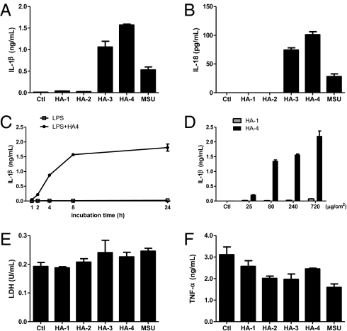
Fig. 1.
Specific forms of HA crystals induce IL-1β and IL-18 secretion from LPS-primed macrophages. (A, B, E, and F) Macrophages from wild-type mice were primed with 50 ng/mL LPS for 14 h and then either left untreated (Ctl group) or stimulated with different forms of HA crystals (240 μg/cm2) or MSU (200 μg/mL) for 8 h. IL-1β (A), IL-18 (B), and TNF-α (F) released into culture supernatants was measured by ELISA. The cytotoxicity of different treatments was determined by lactate dehydrogenase (LDH) release (E). (C) LPS-primed macrophages were stimulated with HA-4 crystals (240 μg/cm2) and culture supernatants were collected at the indicated time of incubation with HA-4. (D) LPS-primed macrophages were stimulated with the indicated amount of HA-1 or HA-4 crystals for 8 h and IL-1β secretion was measured by ELISA. Determinations were performed in triplicate and expressed as the mean ± SEM; several error bars in C are too small to be shown in the graph. Results are representative of at least four independent experiments.

