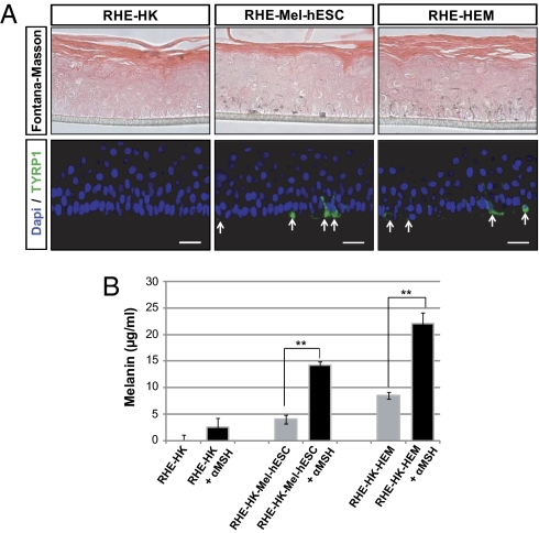
Fig. 4.
Morphological and functional characterization of melanocytes derived from hESCs in an in vitro model of reconstituted melanized epidermis. (A) Fontana–Masson and TYRP1 immunofluorescence staining on reconstituted human epidermis without melanocytes (RHE-HK), containing mel-hESCs (RHE-mel-hESC) or HEMs (RHE-HEM). (Scale bar: 50 μm.) (B) Quantification of melanin content (μg/mL) from reconstituted epidermis containing or not containing melanocytes, without or with stimulation by 1 μM α-melanocyte stimulating hormone (left to right): without melanocytes, with mel-hESCs, and with HEMs. α-MSH, α-melanocyte stimulating hormone. Bars represent the SEM for three experiments (**P < 0.01). All data presented in this figure were obtained in melanocytes during four passages (approximately 50 d) after their isolation.

