
Figure 7.
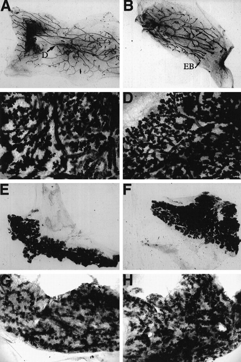
Mammary development is normal in the C/EBPα−/− transplants. Portions of mammary tissue isolated from wild-type (A,C,E,G) or C/EBPα null (B,D,F,H) transplanted glands were fixed and stained with Harris hematoxylin according to standard whole mount procedure. Glands from virgin (A,B), day 13 pregnant (C,D), day 1 lactation (E,F), and day 4 involuted mice (G,H) are included. The whole mounts from 17 days of pregnancy (data not shown) were omitted because they closely resembled the glands isolated at day 1 of lactation. Images of the glands were directly captured from a Sony video camera at 4× (A,B,E,F) or at 10× (C,D,G,H) magnification. Apparent differences in magnification are a result of the original size of each transplant gland. Note the presence of normal ducts (D) in the virgin outgrowths and the presence of terminal end buds in the portion of the ductal tree that has not yet reached the edge of the fat pad in the C/EBPα−/− gland taken at 6 weeks post-transplantation (B).