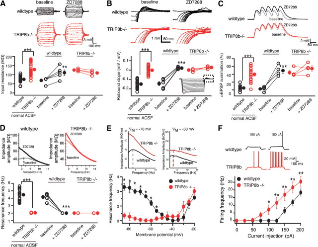
Figure 2.
Deletion of TRIP8b results in functional loss of Ih in CA1 pyramidal neurons. A, Input resistance (RN) was increased in TRIP8b−/− neurons (128.23 ± 6.03 MΩ; n = 30) compared with TRIP8b+/+ (wild-type) controls (74.97 ± 3.4 MΩ; n = 23). B, Neurons from TRIP8b−/− mice had a significantly lower rebound slope (0.00 ± 0.006 mV/mV; n = 30) compared with wild-type controls (−0.14 ± 0.04 mV/mV; n = 23). The dashed box represents the portion of the traces enlarged. C, TRIP8b−/− mice displayed increased temporal summation (39.61 ± 4.31%; n = 18) compared with wild-type controls (4.75 ± 2.05%; n = 11). A–C, Inset, Average response of three traces for 500 ms current injections from −50 to +50 pA in increments of 10 pA (A), 500 ms current injections from 0 to −200 pA in increments of −20 pA (B), and five αEPSP current injections at 20 Hz (C). D, Neurons from TRIP8b−/− mice (n = 28) show no resonance (resonance frequency, fR = 1.04 ± 0.02 Hz) compared with wild-type controls (n = 23; fR = 3.47 ± 0.17 Hz). A–D, ZD7288 (20 μm) significantly altered RN (A), rebound slope (B), temporal summation (C), and fR (D), in wild-type but not TRIP8b−/− neurons. E, Plot of fR versus VM revealed that TRIP8b−/− neurons (n = 5) have a significantly lower fR at hyperpolarized voltages compared with wild-type mice (n = 5) with no significant difference at depolarized voltages. Inset, Impedance amplitude profiles for wild-type (black) and TRIP8b−/− (red) mice measured at −70 mV (left) and −30 mV (right). F, Depolarizing current injection elicited significantly more action potentials from TRIP8b−/− neurons (n = 10) compared with wild-type neurons (n = 8). In all panels, the open symbols represent individual experiments and the filled symbols represent the mean ± SEM. *p < 0.05; **p < 0.01; ***p < 0.001.

