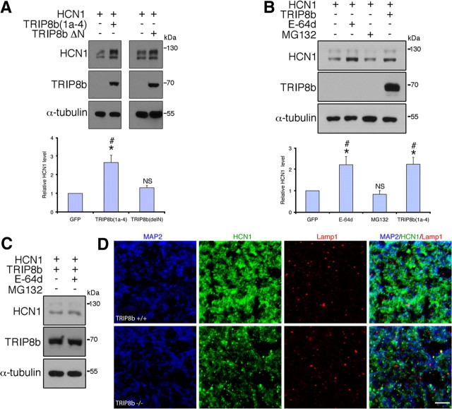
Figure 6.
HCN1 is targeted to lysosomes in neurons lacking TRIP8b. A, Immunoblots from HEK293T cells cotransfected with HCN1 and TRIP8b(1a-4) or TRIP8bΔN demonstrates the TRIP8b N terminus is required for increased HCN1 protein level (*p < 0.05 vs HCN1 only, one-sample t test; #p < 0.05 vs TRIP8bΔN, unpaired t test; n = 5 sets of transfections). NS, Not significant versus HCN1 only. B, HEK293T cells expressing HCN1 and GFP were treated with E-64d (10 μm) or MG132 (10 μm) and HCN1 levels analyzed by Western blot, demonstrating treatment with E-64d enhances HCN1 protein levels similarly to TRIP8b(1a-4) (*p < 0.05 vs HCN1 control, one-sample t test; #p < 0.05 vs MG132-treatment, one-way ANOVA with Tukey's post hoc test; n = 5 sets of transfections). NS, Not significant versus control. C, HEK293T cells expressing HCN1 and TRIP8b(1a-4) were treated with vehicle or E-64d, lysed, and immunoblotted, demonstrating that lysosomal blockade does not further enhance HCN1 protein levels in TRIP8b(1a-4)-expressing cells. D, Confocal images of MAP2 (blue), HCN1 (green), and Lamp1 (red) immunostaining in SLM dendrites reveals increased colocalization of HCN1 and Lamp1 in dendrites of TRIP8b−/− mice versus controls. Scale bar, 5 μm.

