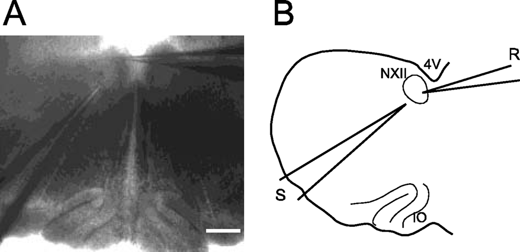
Figure 1.
A- Photomicrograph of the brainstem slice preparation. The fourth ventricle is at the top of the figure; the recording electrode is shown entering from the right side of the field; its tip is located in the left hypoglossal nucleus. A stimulating electrode enters the field from the lower left; the tip is located in the nucleus of Roller. The inferior olive is at the bottom of the slice. Calibration bar: 200µM. B- Diagram of this brainstem preparation. 4V (fourth ventricle), NXII (hypoglossal nucleus), IO (inferior olive), R (recording) and S (stimulating) electrodes.

