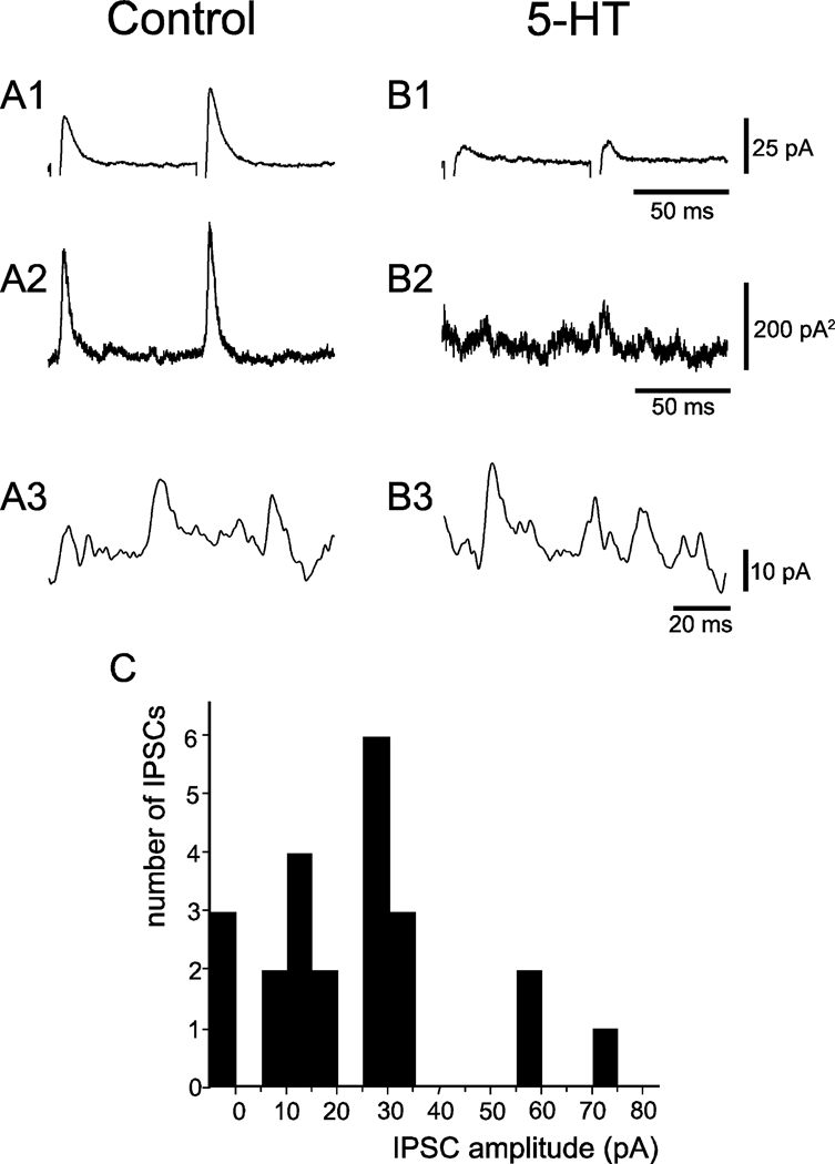
Figure 4.
Presynaptic inhibition of IPSCs by serotonin. A1 and B1, average IPSCs in the same cell evoked by stimuli in a control solution (90 trials) and serotonin-containing solution (43 trials), respectively. Stimulus artifacts have been suppressed. A2 and B2: variance of the current records shown in A1 and B1. A3 and B3: single, high-gain sweeps of spontaneous IPSCs with sizes that are similar to the estimated size of the quanta that comprise the evoked IPSCs in this cell. C: IPSC amplitude histogram from a cell bathed in serotonin showing that 1 to 5 quanta with an estimated size of 14 pA are present in the evoked IPSCs observed in this cell. Note that there were 3 failures in 23 trials.

