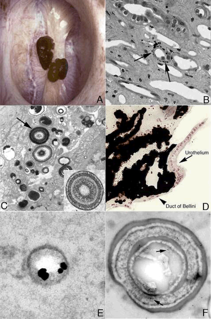
Figure 1. Endoscopic and histologic images of Randall’s plaque in ICSF patients.
Panel A shows two CaOx stones attached to the papilla tip at sites of Randall’s plaque as seen by endoscopy at the time of stone removal. On biopsy (panel B), tissue sections stained by the Yasue method reveals black, small, spherically shaped deposits (arrows) in the basement membrane of the thin loops of Henle, which appears to be the initiating site of plaque formation. By TEM, these deposits appear as multi-layered spheres with alternating light (crystal) and dark (matrix) bands. The smallest deposits were about 50 nm in diameter. Rather dense regions of interstitial plaque located near the papilla tip were commonly noted in ICSF patient (panel D) and at such sites crystal accumulated beneath the urothelium and around the distal ends of ducts of Bellini. Immunoelectron microscopic studies localized osteopontin immunogold particles (dark dots in panel E) at the crystal-matrix boundary whereas the third heavy chain of the inter-alpha trypsin molecule (arrows in panel F) was detected in the matrix layer.

