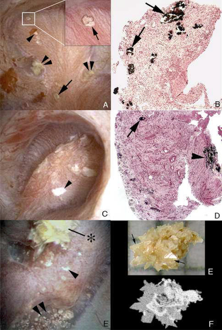
Figure 11. Endoscopic and histologic observations of stone formers with primary hyperparathyroidism.
Endoscopic evaluation of papilla from stone formers with primary hyperparathyroidism shows the coexistence of attached stones (panel A, withinwhite box; , panel E) and plugging of ducts of Bellini (panel A, lower single arrow) on the same papilla. In additions, regions of white plaque (arrowhead, panels A, C and E) and yellow plaque double arrowheads, panels A and E) were also seen on the same papilla. Histopathology of the papillary biopsies showed extensive regions of intratubular plugging of inner medullary collecting ducts and ducts of Bellini (arrows, panels B and D) with areas of interstitial plaque (arrowhead, panel D). Extensive interstitial fibrosis surrounded the plugged tubular segments. Panel E shows an attached calcium oxalate stone (*) before while panel E shows it after detachment. Micro-CT analysis of this same stone (panel F) reveals a mixture of apatite (white regions) and calcium oxalate dihydrate (gray regions).

