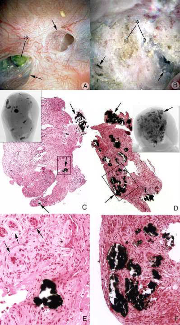
Figure 13. Endoscopic and histologic observations of stone formers with renal tubular acidosis.
Stone patients with renal tubular acidosis had normal papilla apart from an occasional dilated duct of Bellini (arrows, panel A) to severely flattened and fibrotic with pitted appearance (arrow, panel B). These papilla possess multiple dilated ducts of Bellini, some with protruding mineral plugs (*, panels A and B). Histopathlogy and micro-CT imagery shows a range of abnormalities. Plugging of inner medullary collecting ducts varied from minimal in number of tubules involved and size of deposits (arrows, panel C) with some interstitial fibrosis (arrows, panel E), to extensive plugging (arrows, panel D), loss of tubular cells and dense cuffs of fibrotic tissue (arrows, panel F).

