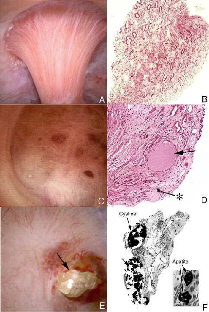
Figure 14. Endoscopic and histologic observations of stone formers with cystinuria.
Papillary morphology varied in these patients with some appearing normal (panel A) to distorted with flattening and greatly dilated opening of ducts of Bellini (panel C). Protruding plugs of cystine were noted in some dilated ducts (arrow, panel E). Histopathology confirmed the observations seen by endoscopy, in that, tissues form papillary biopsies appeared normal (panel B) to abnormal characterized by extensive inner medullary plugging with (panel F) and without mineral deposits (panel D). An occasional mineral plug was noted in loops of Henle (*, panel D). Intraluminal plugs of the ducts of Bellini were primarily cystine in nature while deposits in inner medullary and loops of Henle were always apatite (panel F).

