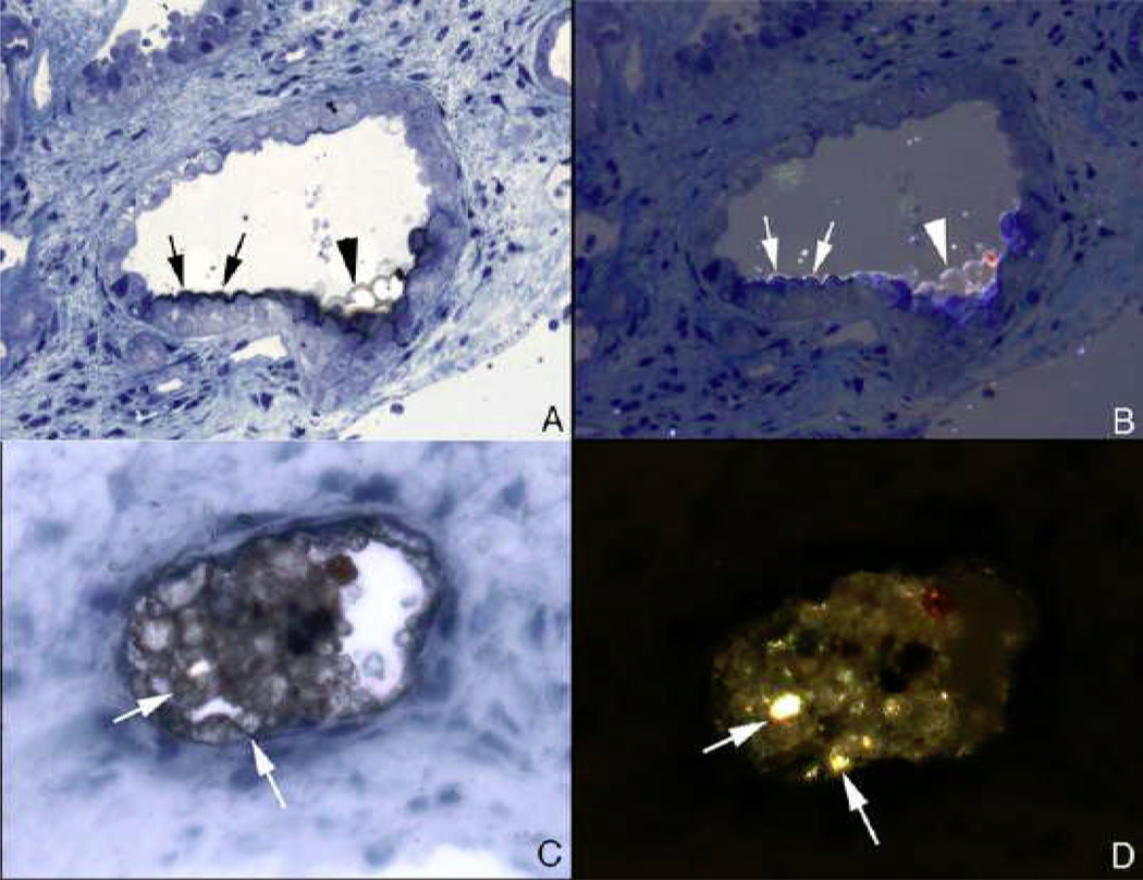
Figure 17. Birefringent and nonbirefringent crystals in inner medullary collecting ducts of stone patients with bypass surgery for obesity.
Panels A and B show serial sections of a inner medullary collecting duct stained by the Yasue method and viewed direct (panel A) and with polarized light (panel B). The inner medullary collecting duct cells seen in panel A are coated with a thin black layer (arrows) that represent areas of calcium deposits. At the arrowhead is a thin region through apical cell surfaces in which the black overlay on the cells is particularly clear. By polarizing light (panel B) the overlay is birefringent suggesting the mineral is calcium oxalate while the rest of the nonbirefringent material is apatite. Panels C and D show another inner medullary collecting duct seen in serial sections by direct (panel C) and polarized light (panel D). The intraluminal plug that partial fills the tubular lumen was stained by Yasue and shows small sites of Yasue negative staining (arrows, panel C) which are birefringent with polarizing optics (arrows, panel D) suggesting the presences of two minerals as seen in panels A and B.

