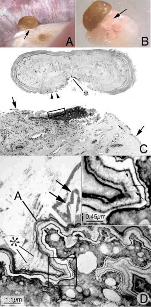
Figure 2. Histologic examination of attached human kidney stone.
During percutaneous nephrolithotomy for stone removal in a ICSF patient, two attached stones were noted on a single papillum (Panel A), a clear region of Randall’s plaque (arrow) is noted at the base of the larger of the two stones. Panel B shows the same stone and the underlying renal tissue with interposing region of Randall’s plaque (arrow) after biopsy removal of this entire complex. The entire biopsy specimen was subsequently decalcified and sectioned first for light microscopy (panel D). During the sectioning process the stone detached from its underpinning so the two images have been approximated in this figure. A large base of interstitial plaque, stained black here by the Yasue method, and framed in part by the rectangle was in continuity with the base of the stone that also reveals a small darkly stained regions (*). This region of plaque is completely devoid of its normal urothelial lining cells. Several attached epithelial cells (arrowheads) were seen at the periphery of the stone while arrows mark the site on the tissue where intact epithelial cells are seen again. The region in Figure 2C marked by the rectangle was prepared and sectioned for transmission electron microscopy (panel D). In this image, the region of interstitial plaque is at the lower right and the stone in the upper left. Between these two regions is a multilayered ribbon structure (labeled A) that forms a sharp boundary. This ribbon is formed by alternating layers of five thin black organic laminia and four, white crystalline laminia (see square and its enlarge in the insert at upper right of panel D). Small arrows in insert show perpendicularly oriented crystals in the white laminia. Various sized crystals (marked by * and large arrows) are seen embedded in the outer matrix layer of the ribbon-like structure.

