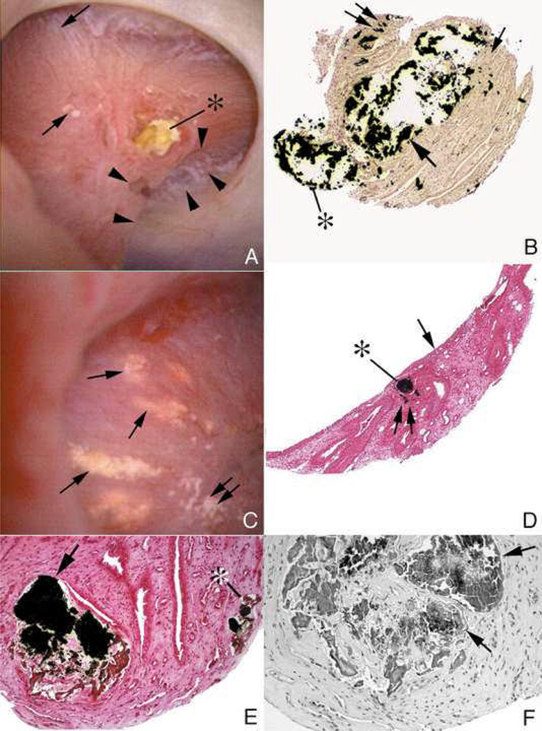
Figure 8. Endoscopic and histologic observations of brushite stone formers.
Brushite stone formers have unique papillary characteristics in that they possess white and yellow plaque as well as protruding plugs from ducts of Bellini. Although the papilla from brushite stone formers have sites of white plaque (arrows in panel A and double arrow in panel C), the most prominent features are mineral plugs protruding from dilated ducts of Bellini (* in panels A and B) and radially oriented sites of yellow plaque (single arrows in panel C) shown to be localized to inner medullary collecting ducts (*, panel D) just beneath the urothelium (arrow, panel D). Many of the papilla of brushite stone formers are deformed as noted by flattening, large dilated opening to ducts of Bellini (Panel A) and large pits (arrowheads in panel A). Histopathology detected grossly dilated inner medullary collecting ducts and ducts of Bellini filled with mineral deposits (arrows in panel B) shown to be apatite. These dilated tubules were surrounded by interstitial fibrosis. Regions of interstitial plaque were easily found (double arrows, panels B and D). Using light microscopy of 1 micron thick plastic sections of decalcified papillary biopsies, extensive regions of cellular damage with mineralization were seen in inner medullary collecting ducts (arrows, panels E and F) and loops of Henle (*, panel E) adjacent to normal appearing tubular segments (panel E). Extensive interstitial fibrosis is noted around these sites of tubular injury.

