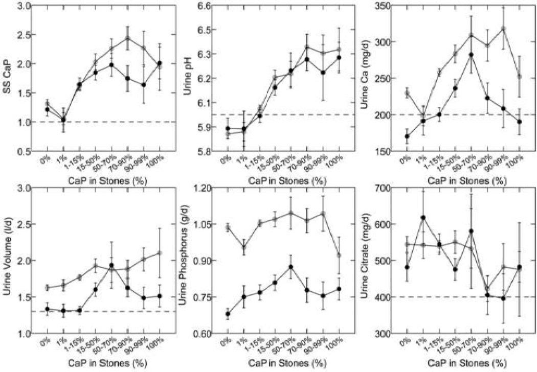
Figure 9. Urine measurements in ICSF with increasing stone calcium phosphate (CaP) percent.
With increasing percent of CaP in analyzed stones (X-axes of all panels) SS CaP and urine pH (upper left and middle panels) rose progressively, and urine calcium excretion (upper right panel) in a less constant manner. Urine volume, and phosphate and citrate excretions (lower panels) showed no consistent relationship to stone CaP.

