Figure 2.
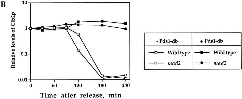
Pds1p’s ability to inhibit Clb2p degradation is independent of the spindle damage checkpoint protein Mad2p. Wild-type, GAL1–pds1-db, mad2-1, and mad2-1 GAL1–pds1-db cells (strains VG906-1A, VG1369, VG1346-10D, and OCF2101, respectively), were grown in YPR and arrested in HU as described in the legend to Fig. 1. The expression of GAL1–pds1-db was induced for 30 min, after which the cells were released from the HU arrest into fresh YPRG medium containing the α mating factor, to inhibit cell cycle progression in the following G1. Samples were taken at the indicated time points and processed for Western blot analysis (A,B) or cell morphology (C), as described in Materials and Methods. (B) The relative Clb2p levels as detected by Western blot analysis (A) in the different strains, as indicated. (C) The distribution of cell types in the mad2-1 and mad2-1 GAL1–pds1-db cultures as scored by DAPI staining. The percentage of large budded, single nucleated cells (squares) or anaphase cells (circles), in the absence (open symbols) or presence (solid symbols) of Pds1-db, are shown.


