
Figure 1.
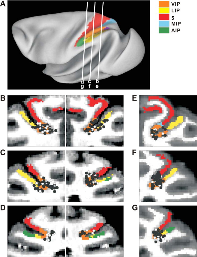
Anatomical localization of recording sites. A, Inflated cortical surface illustrating the locations of the coronal sections drawn in B–G. B–D, Coronal sections from both hemispheres of monkey J, spaced 4 mm apart, are shown from posterior (B) to anterior (D). E–G, Coronal sections from the right hemisphere of monkey C are shown from posterior (E) to anterior (G). Cells located within 2 mm of each section were projected onto that section. The black symbols represent single units with significant tuning to either vestibular or visual translation. The white symbols represent cells that showed no directional tuning.