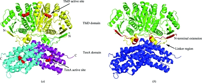
Figure 2.
Stucture of S. cerevisiae THI20. (a) A ribbon diagram illustrating the structure of the THI20 dimer with each domain colored separately (ThiD-like domains colored yellow and green and TenA-like domains colored cyan and purple). The locations of the active sites are shown with a space-filling model of HMP and are indicated by arrows. (b) A model of THI20 showing the regions with no sequence similarity to other ThiD or TenA proteins. The linker region joining the ThiD-like domains (green) and the TenA-like domains (blue) is shown in yellow, while the N-terminal extension is shown in red.

