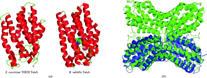
Figure 5.
Comparison of the overall structure of the THI20 TenA-like domain to B. subtilis TenA. (a) The THI20 TenA-like protomer is shown alongside the protomer of B. subtilis TenA (PDB entry 1yak). In both cases helices are colored red and loop regions are colored green. The active site of B. subtilis TenA is indicated by HMP, which is shown as a space-filling model with green C atoms, red O atoms and blue N atoms. (b) A superposition of the THI20 TenA-like dimer (blue) with the B. subtilis TenA tetramer (green).

